
Learn Your Brain
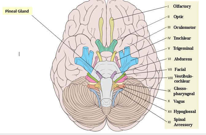
There are twelve cranial nerves which originate in the brainstem, which are identified by Roman numerals (as well as a common mane) in order as they arise from progressively lower portions of the brainstem or forebrain. The functions are varied, and include control of movement or the eyes, hearing, sensation, smell, sight, mucous membrane function, and control of bowel, heart and lung functions.
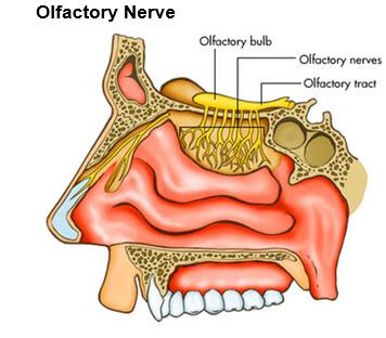
Cranial nerve I, known as the olfactory nerve, conveys the sense of smell. Within the mucosa of the nasal cavity exist fine nerves, which ascend through the cribriform plate (part of the skull base which contains fine perforations through which nerves may travel) and then travel in the olfactory tract to enter the undersurface of the brain.
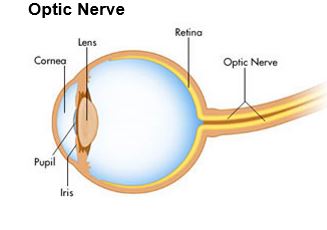
The optic nerve (cranial nerve II) provides for vision. Each of the two optic nerves leave the orbit through the optic foramen and unites with the other optic nerve at the optic chiasm. In the chiasm, some of the nerve fibers cross to the other side. The optic tract continues and sends signals to the lateral geniculate body, then through the optic radiations to the calcarine cortex of the occipital lobe (where vision is perceived).
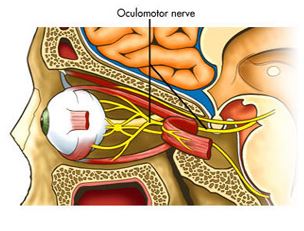
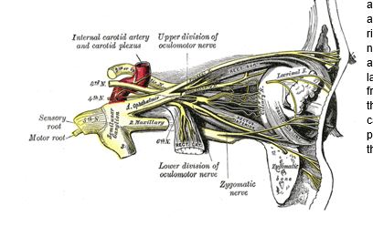
The nucleus for the occulomotor nerve (cranial nerve III) lies in the upper portion of the brainstem. This nerve controls certain extraoccular muscles and movements. The Edinger Westphal nucleus is part of the third nerve nucleus, and sends parasympathetic fibers to cause constriction of the pupils. Through a sophisticated connection of fibers in the brainstem, the occulomotor nerve (cranial nerve III) interacts with the abducens nerve (cranial nerve VI), causing the eyes to move in unison, allowing stereo vision. Of note is that the parasympathetic fibers (part of the autonomic nervous system) cause constriction of the pupils, while the sympathetic fibers (also part of the autonomic nervous system) cause dilation of the pupils.
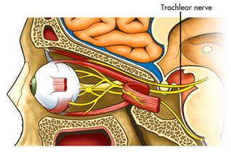
The fourth cranial nerve, known as the trochleear nerve, supplies the superior oblique muscle of the eye. The nucleus for this nerve lies in the midbrain, and passes through the cavernous sinus to enter the superior orbital fissure of the orbit.
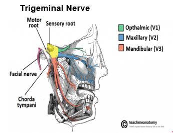
The fifth cranial nerve, known as the trigeminal nerve, is involved with both sensation to the face as well as the muscles of mastication. Both the motor and sensory portions of the trigeminal nerve emerge from the lateral aspect of the pons. The nerve travels out to the Gasserian ganglion, from where three branches or divisions of the nerve are given off. The ophthalmic division supplies sensation to the forehead and cornea. This division is responsible for the corneal reflex (when something touches the cornea, the eye reflexively blinks shut). The maxillary division supplies the cheek, and the mandibular dividion, the jaw. A division of the mandibular branch supplies the muscles of mastication (masseter, pterygoids and temporalis), and also supplies sensation to the cheek and gums through the buccal nerve. The lingual branch helps in supplying taste to the anterior two thirds of the tongue.
The sixth cranial nerve is also known as the abducens nerve, as it is responsible for abducting (pulling the eye away) the eye. The right eye will look to the right, away from the nose and the body, as a result of the abducens nerve causing constriction of the lateral rectus muscle. The nerve leaves the front of the brainstem between the pons and the medulla. It then travels through a dural canal known as Dorello’s canal, before it passes through the cavernous sinus, to enter the orbit
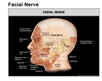
The seventh cranial nerve is also known as the facial nerve. It carries mainly motor fibers to the muscles of facial expression, but it also carries fibers which relay taste, as well as fibers carrying parasympathetic information. The motor nucleus lies in the lower pons, and the facial nerve exits the lateral aspect of the brainstem, to enter the internal auditory canal. The parasympathetic component (goes to the lacrimal gland to produce tears) and the taste fibers (receive taste from the anterior two thirds of the tongue, through the chorda tympani nerve) both run in a small nerve adjacent to the facial nerve, known as the nervus intermedius.
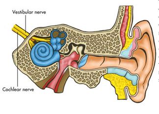
The eight cranial nerve (also known as the vestibulocochlear nerve) consists of two portions, the vestibular portion and the cochlear portion. The vestibular portion is responsible for responding to rotational and linear acceleration, which helps maintain equilibrium and body orientation in space. Semicircular canals contain endolymph, which during movement, displaces small hair cells, which in turn send a signal through the vestibular nerve. The cochlea is responsible for hearing, as it converts sound waves into signals in the cochlear neurons.
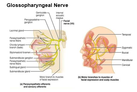
The ninth cranial nerve (known as the glossopharyngeal nerve) serves a combination of motor, sensory and parasympathetic function. The stylopharyngeus muscle is supplied by it. The parotid gland is supplied by it. Taste from the posterior third of the tongue is also supplied by this nerve. The nerve leaves the medulla portion of the brainstem, as 5 to 6 small rootlets.
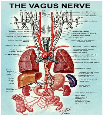
The tenth cranial nerve (known as the vagus nerve) provides motor, sensory and parasympathetic function. The motor function supplies the pharynx, soft palate and larynx, and originates in a nucleus in the brainstem known as the nucleus ambiguus. The parasympathetic function supplies the thoracic and abdominal viscera. Sensory fibers arise in the pharynx, larynx and external auditory meatus.
The vagus nerve leaves the brainstem as a number of converging rootlets, and leaves the skull through the jugular foramen.
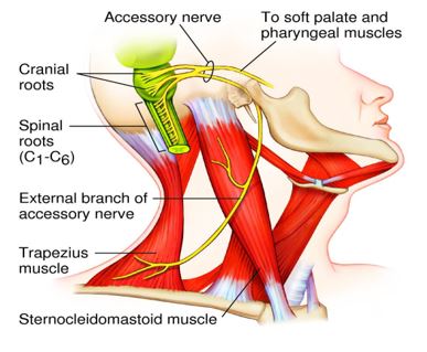
The eleventh cranial nerve (known as the accessory nerve), originates in the medulla or the brainstem, as well as the upper to mid cervical spinal cord. The spinal portion arises to enter the skull through the foramen magnum, and it joins with the cranial portion to exit the skull through the jugular foramen. These nerves supply motor function to the sternocleidomastoid and trapezius muscles.
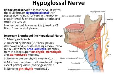
The twelfth cranial nerve (known as the hypoglossal nerve) supplies the intrinsic muscles of the tongue. The nerve originates in the hypoglossal nucleus in the medulla of the brainstem, and leaves the skull through the hypoglossal canal.